Cancer detection through EUS technology
10 August 2019
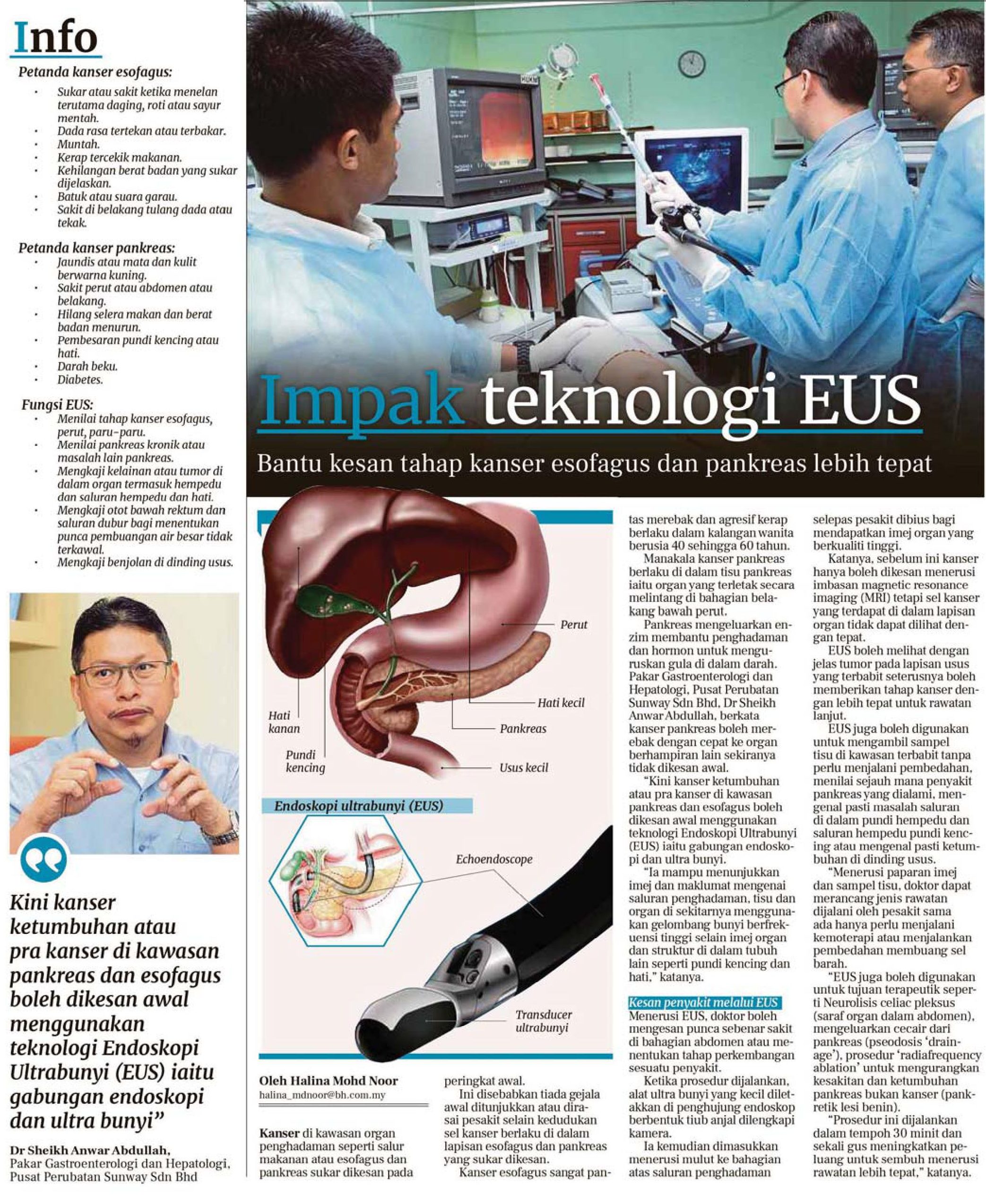
Cancer in digestive organs such as the gastrointestinal tract or oesophagus and pancreas are difficult to detect at an early stage. This is because there are no initial symptoms shown or felt by the patient other than the cancer cells occurring in the lining of the oesophagus and pancreas which is difficult to detect.
Oesophageal cancer is rapid and aggressive, and often occurs in women aged 40 to 60 years. Meanwhile, pancreatic cancer occurs in the pancreatic tissue which is an organ located horizontally in the lower back of the stomach. The pancreas releases enzymes that help digestion and hormones to manage blood sugar levels.
Sunway Medical Centre Consultant Gastroenterologist and Hepatologist, Dr Sheikh Anwar Abdullah said pancreatic cancer can spread quickly to other nearby organs if not detected early.
“But now cancer tumours or pre-cancer in the pancreas and oesophagus can be detected early using the endoscopic ultrasound (EUS) technology, which is a combination of an endoscopy and ultrasound.
“It is able to show images and information from the digestive tract and tissues and organs around it using high frequency sound waves as well as images of organs and structures in other bodies such as the bladder and liver,” he said.
Through EUS the doctor can detect the real cause of pain in the abdomen or determine the stage of a disease.
During the procedure, a small ultrasound device is placed at the end of an elastic tube-shaped endoscope equipped with a camera. It is then inserted through the mouth into the upper part of the digestive tract after the patient is anesthetised to obtain high quality organ images.
Dr Sheikh said before this cancer can only be detected through a magnetic resonance imaging (MRI) scan but cancer cells found in the layers of an organ cannot be seen accurately.
EUS can clearly see tumours in the lining of the intestine which in turn provides the stage of cancer levels to be determined more accurately for further treatment.
EUS can also be used to take tissue samples in the affected area without having to undergo surgery, assess the extent of pancreatic disease, identify problems in the gallbladder and gallbladder ducts, or identify tumours in the intestinal wall.
“Through the display of images and tissue samples, doctors can plan the type of treatment whether the patient only needs to undergo chemotherapy or a cancer cell removal surgery.
“EUS can also be used for therapeutic purposes such as neurolysis celiac plexus (nerve organ in the abdomen), removal of fluid from the pancreas (pseudocyst drainage) and radiofrequency ablation procedure to reduce pain and non-cancerous pancreatic growth (pancreatic lesion benign).
“This procedure is carried out within 30 minutes and increases the chances of recovery through more accurate treatment,” Dr Sheikh said.
Source: Berita Harian
Back
