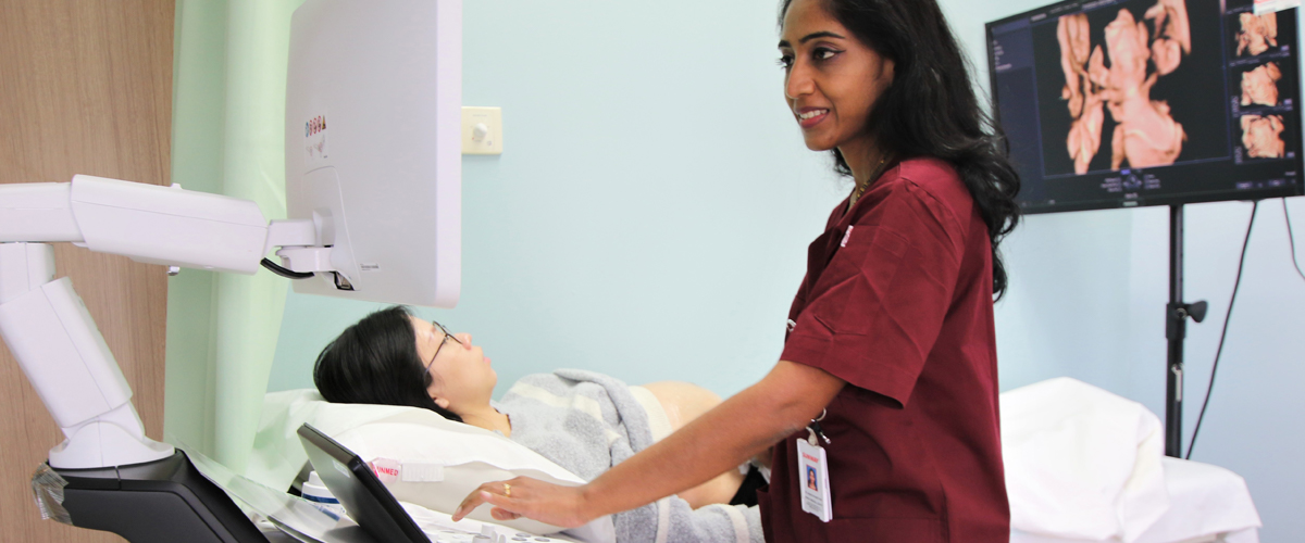
Prenatal Ultrasound

  • Home
  • >
  • Prenatal Ultrasound

The developing fetus is a source of immense joy. Prenatal ultrasound assesses fetal wellbeing. While the vast majority of babies are born without birth defects, there is always a risk of abnormal fetal development, regardless of parents’ age, family history, maternal medical condition, or lifestyle habits.

Congenital abnormalities and birth defects affect 2% to 3% of all pregnancies, but most of them are minor. Very few babies have major birth defects. That is why having a scan is not just a joyful family event, but an important procedure to shed light on the health of the fetus.

Anomaly Scan

The anomaly scan (also called morphology scan, anatomy scan, or 20-week scan) is a detailed ultrasound scan in 2D image that looks at your baby’s body and identifies the position of the placenta, the umbilical cord, the amniotic fluid around your baby, your womb, and your cervix.

It enables the doctor to assess if the baby has any abnormalities as it grows, by looking at its brain, face, heart, lungs, stomach, abdomen, kidneys, spine, and limbs. The gender can also be identified with a high level of accuracy.

While major anomalies involving organ systems can generally be identified, some minor defects may remain undetected and certain types of anomalies are not amenable to be detected before birth.

The benefit of this scan is that if there are no abnormalities detected, parents and doctors can be assured that the baby is physically normal. If any issues are detected, counselling and further tests will need to be carried out, so that parents can make informed decisions regarding the management of their pregnancies.

This scan is ideally carried out between 20 to 24 weeks of pregnancy. However, it can be done as early as 18 weeks and as late as 28 weeks of pregnancy.

Conditions that can be detected during an anomaly scan include :

  • Spina bifida
  • Anencephaly (absence of a part of the brain and skull)
  • Cleft lip
  • Gastroschisis (intestines growing outside the body)
  • Serious heart conditions
  • Limb defects, etc.

3D SCAN

If the baby’s position is optimal during the anomaly scan, a 3D scan may also be carried out.

3D ultrasounds allow parents to have a more life-like view of the baby’s face, and can be exciting to watch.

4D SCAN

While 3D ultrasounds produce clear still images of the baby, 4D ultrasounds take it one step further by showing the baby in video form. Parents get to see the baby in live-action— smiling, smirking, thumb-sucking, yawning, and more.

These scans are ideally carried out between 26 to 34 weeks of pregnancy.

3d scan

3D SCAN

If the baby’s position is optimal during the anomaly scan, a 3D scan may also be carried out.

3D ultrasounds allow parents to have a more life-like view of the baby’s face, and can be exciting to watch.

4D SCAN

While 3D ultrasounds produce clear still images of the baby, 4D ultrasounds take it one step further by showing the baby in video form. Parents get to see the baby in live-action— smiling, smirking, thumb-sucking, yawning, and more.

These scans are ideally carried out between 26 to 34 weeks of pregnancy.

General Scanning Timeline

8 - 14
weeks:
Dating
Scan
  • Confirm baby’s due date
  • Detect baby’s heartbeat
  • Rule out ectopic pregnancy (pregnancy outside of the uterus)
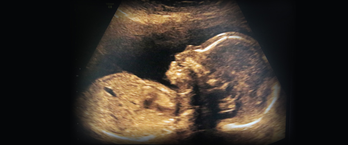
11 - 13
weeks:
First Trimester
Scan
  • Measure nuchal translucency (NT)
  • Screen for risks of Down syndrome and other major trisomies, i.e. Edward and Patau syndromes by First Trimester Screening (FTS) or Noninvasive Prenatal Testing (NIPT)
  • Screen for preeclampsia risk
20 - 24
weeks:
Anomaly
Scan
  • Rule out several fetal and placental conditions
  • Confirm normal physical and anatomical features within the limits of the scan
  • Identify gender
  • Measure cervical length to predict risks of preterm birth
  • Measure uterine blood flow to evaluate risks of preeclampsia and growth restriction
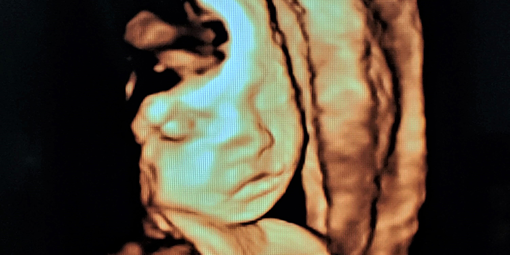
25
weeks
onwards
  • Obtain good 3D and 4D ultrasound images of the baby
32 - 36
weeks:
Third
Trimester
Scan
  • Detect baby’s position, i.e. whether head is down, bum first (breech), or lying sideways (transverse), and assess fetal growth
  • Check amniotic fluid volume and blood flow between baby and the placenta

Frequently Asked Questions

What are the differences between 2D, 3D, and 4D ultrasound ?

2D ultrasound is the traditional screening method that provides outlined, 2-dimensional, flat images of the baby, which usually is performed and interpreted by a professional or trained sonographer.

3D and 4D ultrasounds are able to give solid, life-like images that parents can identify, giving them a chance to connect with the baby much earlier. They also provide clearer pictures for doctors to show the parents if there are any problems with the baby.

4D ultrasounds are even more realistic by showing the baby’s movement, so parents can watch the baby kick or smile in real time.

Are they different from a prenatal diagnostic exam ?
Are ultrasounds safe ?
How long does an ultrasound session take ?
How should I prepare myself before an ultrasound ?
When will I get the results of my ultrasound ?
Who should I look for to get a first trimester scan, 2D anomaly scan, or a 3D/4D ultrasound ?